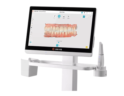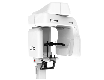
DEXIS™ OP 3D LX (POA)
Dexis
The next generation of DEXIS cone beam technology
Built on OP 3D technology, this multimodality imaging platform expands your 3D diagnostic capabilities with a wide range of clinical applications that support your evolving practice and enhance diagnostic confidence.
Built to increase practice efficiency
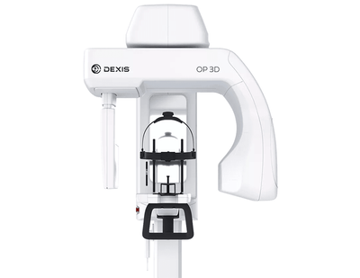
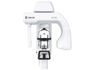
The 2D and 3D imaging options built into the ORTHOPANTOMOGRAPH™ OP 3D™ LX unit cover a full spectrum of dental extraoral needs, from endodontics to complex surgical cases. This next generation system offers flexible field of view (FOV) options ranging from 5 (H) x 5 (D) cm up to 15 (H) x 20 (D) cm – which is the largest view option available on a DEXIS OP 3D platform to date. With shorter scan times, the OP 3D LX captures the maxillofacial complex and large diagnostic areas in one non-stitched scan for fast workflows.
One versatile imaging platform
Flexible volume sizes
With the largest sensor on a DEXIS OP 3D platform, OP 3D LX offers flexible FOV options ranging from 5x5 quadrant scans up to 15x20 complete maxillofacial complex high-resolution scans.
A scalable solution
Easily upgrade your system to meet your evolving diagnostic needs with adding larger volume sizes or the cephalometric modality.
High-quality images
Expand your diagnostic confidence and capabilities with the artifact and noise reduction filters embedded into the system software that give you the consistent clear high-quality images.
Explore the expanded view options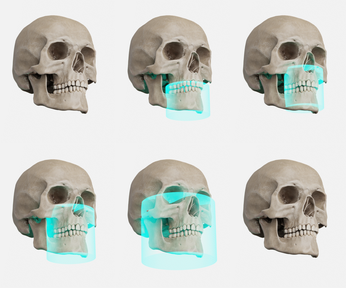
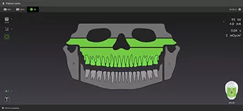
User interface
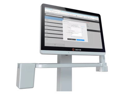
OP 3D LX offers an intuitive user interface that enables you to easily position the patient, visually choose the areas of interest with your 3D, panoramic or cephalometric settings and preview the X-ray image shortly after exposure without opening any image viewing software.
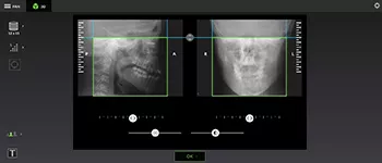
Intuitive accuracy
The intuitive user interface in the OP 3D LX system makes it easy to select your field of view and allows for accurate anatomy visualization, vertical adjustments, and bi-directional scout modifications to capture only structures of interest.
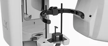
Re-engineered head support
The new head support design provides options to scan the patient without interfering with the patient’s soft tissue profile optimized for orthodontic and surgical applications.
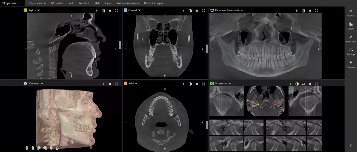
Stitch-free scans
You can accurately diagnose, plan, and treat your patients with confidence using single pass capture with no stitching on every scan size.
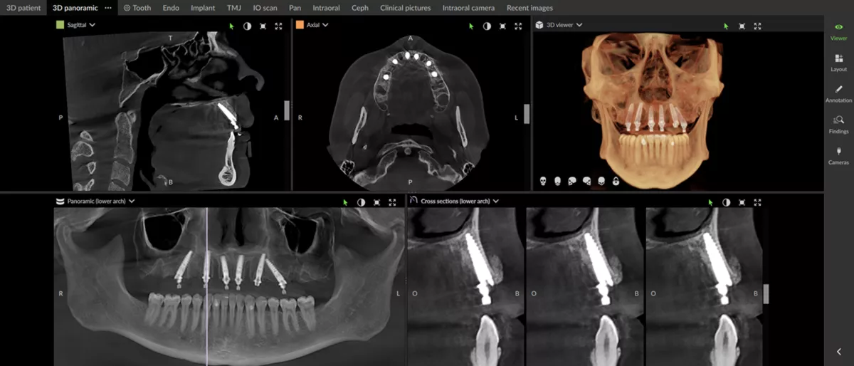
Enhanced visibility
Enjoy the next generation of automated ICE* (Implant Contrast Enhancer) and MAR (Metal Artifact Reduction) to provide greater visibility of internal metal structures of existing implants, while minimizing the impact of metal and restorations.

Cloud-based service connectivity
This OP 3D LX feature simplifies service and maintenance for improved practice productivity and uptime.
Assisted intelligence for workflow efficiencies
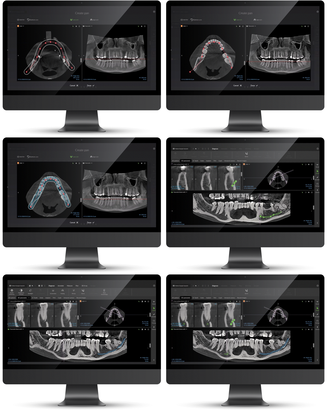
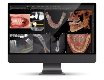
Our intuitive award-winning software features, in DTX Studio™ Clinic, support a more efficient workflow allowing you to spend less time in the software and more time with your patient. Some of the many assisted intelligent features include:
- Automatic focal trough detection
- Patient positioning correction
- Mandibular nerve canal annotation
